Visualizing New Frontiers

Scientific American Custom Media talks to 2020 Kavli Prize Laureate Ondrej Krivanek about how breakthroughs in microscopy drive discovery.
Biggest Questions In Science
Living Paramecium photographed through a microscope. The outline is blurred by the movement of the cilia

Finding focus

From the 17th-century observations of Robert Hooke and Antonie van Leeuwenhoek to the 19th-century observations of cellular organelles and the nervous system, biology advanced as microscopy improved. But then the technology ran into physical limitations. In 1873 German physicist Ernst Abbe discovered that the wavelength of the illuminating light limits the resolution of light microscopy to about 200 nanometers. Many important objects — including viruses, ribosomes, and cell membranes — were too small to be visualized.

Credit: Library Book Collection/Alamy
Professor Ernst Ruska (1906-1988) photographed  at the Max Plank Institute for Physics in Berlin

Electron microscopy emerges

By the 1920s, physicists had discovered that electrons, like photons of light, behaved as both particles and waves — and that their wavelength was short enough, in theory, to visualize objects as small as atoms. This drove Ernst Ruska (shown here) and Max Knoll to build the first electron microscope in 1931. By visualizing objects with electron beams rather than light, electron microscopes clarified the microscopic structure of bacteria, viruses, materials and minerals. This gave scientists a new tool to understand nature.

Credit: Thomas Hollyman/Science Photo Library

Intricate images

Throughout the 20th century, the two main variants of electron microscopy — transmission electron microscopy and scanning electron microscopy — revealed the microscopic structure of bacteriophage, poliovirus, pollen, chalk, human cells (shown) and hundreds of other objects. Yet the method failed to live up to its potential. Like a person with an astigmatism, the magnetic lenses in electron microscopes could not focus the electron beam well enough to resolve the tiniest objects, such as molecules or atoms. This problem, known as aberration, remained unsolved for 60 years.

Credit: Enterobacteria Phage T4: Science History Images/Alamy. Polio virus: Joseph J. Esposito; F. A. Murphy/CDC. Pollen: Dartmouth College Electron Microscope Facility. Calcareous phytoplankton: Steve Gschmeissner/SPL/Getty. Human lymphocyte: Science History Images/Alamy
Rose-EELS focussing electron beam graphic

Correcting aberration

In 1990, physicist Harald Rose figured out how to correct for aberration and give the electron microscope “glasses,” as he put it. Physicists Maximilian Haider and Knut Urban then fit a transmission electron microscope with those glasses. Meanwhile, physicist Ondrej Krivanek built an aberration corrector (shown here) for the scanning transmission electron microscope. Aberration correctors use a stack of donut-shaped magnetic lenses to shape and reshape a wayward electron beam and train it precisely on the sample. The work earned the four physicists the 2020 Kavli Prize in Nanoscience for helping scientists directly visualize atoms.

Credit: Falconieri Visuals
Ondrej installing the Nion MarkII aberration corrector on the SuperSTEM1 instrument in Cambridge, prior to the microscope being move to the SuperSTEM Laboratory

Steering the beam

Before Krivanek designed an aberration corrector for the scanning transmission electron microscope, senior scientists told him that he would fail and the project would bury his career. But Krivanek and his partner, engineer Niklas Dellby, persevered and succeeded, and then went on to cofound the Nion Company to build advanced electron microscopes. For the first time, scientists could resolve common elements like carbon and nitrogen at the atomic level.

Watch Ondrej Krivanek talk about getting hooked on electron microscopy. Credit: Uwe Falke/SuperSTEM
HeLa cells with Adenovirus, SEM
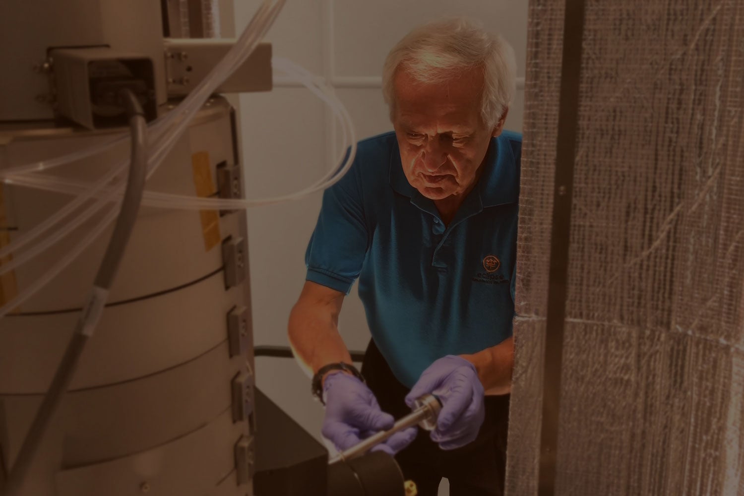
“The world is made of atoms. They are the building blocks, and everything is made out of them. We are made of them. The air that we are breathing is made of them. The coronavirus is made out of atoms. And if you know how the building blocks come together, you know a lot about the world.”
00:19
Listen to the full podcast

Atoms in focus

Localizing atoms was critical, but it was equally important to chemically identify them. In 2010 Krivanek and colleagues reported in Nature that they had done both simultaneously. They used a high-resolution, aberration-corrected scanning transmission electron microscope that Nion had built to localize each atom in a hexagonal crystal of boron nitride, and identify which chemical element it was. This allowed them to spot the boron (red) and nitrogen (green) atoms, as well as impurities of carbon (yellow) and oxygen (blue).

Credit: Krivanek, O. et al. Nature https://doi.org/10.1038/nature08879 (2010).
Double cone figure

Spotting the smallest atom

As aberration-corrected electron microscopes improved, scientists began visualizing lighter and lighter atoms. In 2020, researchers peered into a material called a metal-metal hydride and visualized the smallest atom, hydrogen, for the first time. To do so, they used a scanning transmission electron microscope and a new visualization method called integrated differential phase contrast, they reported in Science Advances. Visualizing hydrogen atoms should help scientists grasp how they behave in solids, and how external forces, such as mechanical stress, alter that behavior. This knowledge is essential for new energy applications, such as storing hydrogen in fuel cells.

Credit: Reprinted with permission of AAAS from de Graaf et al (2020). Resolving hydrogen atoms at metal-metal hydride interfaces. Sci. Adv. 6 (5) eaay4312. © The Authors, some rights reserved; exclusive licensee AAAS. Distributed under a CC BY-NC 4.0 License

Good vibrations

The latest scanning transmission electron microscopes do more than localize and identify atoms. Krivanek and his colleagues at Nion have added a form of spectroscopy to these microscopes that detects how atoms vibrate and interact with their neighbors. The method, called vibrational electron energy-loss spectroscopy (vEELS), can help solid-state physicists predict how sound and heat will move through a material. This, in turn, can help engineers design and build better thermal insulators and thermoelectric devices, which convert heat into electricity.

Credit: Demie Kepaptsoglou/SuperSTEM
Structure - GABAAR reconstruction

Frozen treasure

Scientists have used x-ray crystallography for half a century to visualize the atomic structure of proteins, the workhorses of our cells. But the method is effective only for proteins that form crystals, and many important proteins do not. In 2020, researchers reported in Nature that a different method could produce atomic-resolution images of proteins in frozen tissue samples. The method, called cryo-electron microscopy, could help researchers visualize biologically critical proteins, which could lead to new drugs that target them to fight disease.

Credit: Nakane, T. et al. Nature https://doi.org/10.1038/s41586-020-2829-0 (2020)
Dr. Kimani working in his lab fabricating and characterizing perovskite solar cells  based on triple-cation and all-inorganic absorber chemistries for the upcoming MISSE-15 launch to the International Space Station by NASA

Spray-on solar cells

Visualizing the atomic structure of inorganic compounds such as lithium or silicon can help materials scientists understand the mechanical and electrical properties of a material by analyzing its atomic structure. Insights from such experiments could improve the energy-conversion efficiency and reduce the cost of gallium-arsenide layers, leading to transformative technologies, such as thin-film solar photovoltaics.

Credit: Dennis Schroeder/NREL
Ondrej Krivanek loading samples into the scanning transmission electron microscope
“The way I tend to think about these things is, Here's a powerful experimental capability that should be available if we sweat the details, so let's build it and then let's see what we can do with it. And let’s make it available to the world and see what people do with it in different laboratories. It's a model that's worked very well.”
00:24
Listen to the full podcast
This article was produced for The Kavli Prize by Scientific American Custom Media, a division separate from the magazine's board of editors. To learn more about Kavli Prize-winning research, see kavliprize.org. Share The Project Credits: Fruit fly images by Power and Syred/Science Photo Library. HeLa cell image by Science History Images/Alamy. Ondrej Krivanek image courtesy of Nion Co.